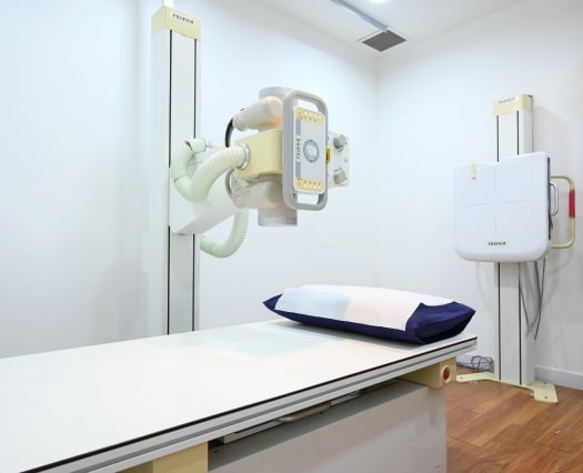
General X-Rays
A chest X-ray is an imaging test that makes use of small amounts of radiation to produce pictures of the tissues, bones, and organs of the body. When used on the chest, it is used to spot diseases or abnormalities of the airways, bones, heart, lungs, and the blood vessels.
An abnormal chest X-ray reading might indicate there is a possible problem in the chest. A chest X-ray can be ordered for various reasons. For instance, it can be used to assess injuries resulting from an accident or monitor the progression of a certain condition like cystic fibrosis.
Any abnormal chest X-ray finding will be looked into to identify any underlying conditions so it can be addressed accordingly. A chest X-ray is quick, easy and effective. It has been used for decades now to help doctors view some of the body’s most vital organs.
Preparation
Before the examination
- Different examinations have specific preparation requirements. Please check with your doctor/ nurse if you are unsure.
- If you have taken similar examinations in the past, it is advisable to bring them along on your appointment.
- When making your appointment please inform the nurse if you are on medications for conditions like asthma, diabetes, cardiac or kidney diseases etc, or if you think you may be pregnant.
During the examination
- When your appointment is due, you will be led to the examination room by the radiographer.
- You will then be requested to position yourself either on the examination bed or to remain standing when the examination is conducted. This will be explained clearly to you by the radiographer who will be conducting the examination. At no time should you be feeling uncomfortable and if you are, do inform the radiographer immediately.
- Depending on the type and complexity of the examination, each examination will usually take between 10 – 35 minutes.
After the examination
- The collection of results will depend on your doctor’s request. The result will either be collected by you where you will bring it back to your doctor for further consultation or it will be dispatched to your doctor who will then schedule an appointment to discuss the findings with you.
FAQ
Doctors may order a chest X-ray if they suspect the symptoms that manifest are connected to possible problems in the chest. Some of the suspicious symptoms to look out for can include:
- Fever
- Persistent cough
- Chest pain
- Shortness of breath
The mentioned symptoms can be the result of some of the following conditions:
- Emphysema
- Lung cancer
- Broken ribs
- Pneumonia
- Heart failure
- Pneumothorax
Chest X-ray is also used to check the shape and size of the heart. Any abnormalities in the size and shape can indicate any possible problems with the heart function. Doctors also sometimes use chest X-rays to monitor the progress after any surgery done to the chest area.
Doctors can also use chest X-rays to see if any implanted materials are still in place. It is also used to ensure the patient is not experiencing any fluid buildup or air leaks.
Chest X-rays will require very minimal preparation. Prior to the procedure, patients will be asked to remove any body piercings, eyeglasses, jewelry, and other metals the individuals may be wearing.
Patients with any implanted device like pacemaker or heart valve should tell the individual performing the procedure. It is possible that you can still go through with the procedure even with metal implants. However, other scans like MRIs may be risky for those who have metal in their bodies.
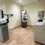
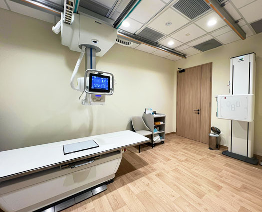
X-ray is carried out in a special room with a movable camera that is attached to a metal arm. You will be standing next to a “plate.” The plate contains a special sensor or an X-ray film that records the images taken on a computer.
A front and side view of the chest will be taken. While the images are taken, you need to hold your breath so the chest remains completely still. Any movement while the images are taken might turn out blurry.
As the radiation passes through the body, bones, muscles of the heart, and other dense materials will appear white. After the images have been captured successfully, the procedure is complete and you can change back into your clothes and go about your day. The whole process will only take around 20 minutes from start to finish.
X-rays are not recommended for pregnant women as the radiation might cause harm to the unborn baby. If you suspect you are pregnant, it is recommended that you tell your doctor first before agreeing to a chest X-ray.


